The Standard Views
Parasternal, apical, subcostal, suprasternal — five windows, twelve views.
Acoustic Windows and Doppler Alignment
Each standard transthoracic window exploits a region where the ultrasound beam avoids lung (air) and rib (bone), both of which scatter and attenuate the signal; the parasternal window uses an intercostal space adjacent to the sternum, the apical window uses the point of maximal impulse, and the subcostal window transmits through the liver as an acoustic pathway. Higher-frequency transducers improve axial resolution but penetrate less deeply, which is why adult transthoracic imaging typically uses 2-4 MHz. Doppler measurements additionally require the ultrasound beam to be aligned as parallel as possible to the direction of blood flow, which is why apical views are preferred for Doppler interrogation of LVOT and mitral inflow, while parasternal views are preferred for 2D structural detail.
Parasternal Long Axis
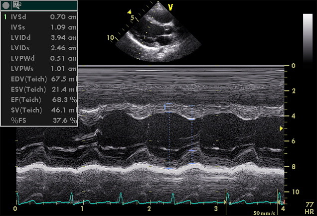 LV, LA, LVOT, aortic and mitral valves. Measure LV wall thickness, chamber diameter, LA size, aortic root, valve mobility.
Parasternal Short Axis
Apical 4-Chamber
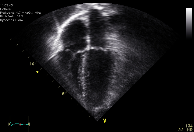 All four chambers, MV and TV, both septa. Foreshortening is the main pitfall — apex must be visible.