Back to curriculum
Domain

Adult Congenital Heart Disease

Modules

Atrial Septal Defect

The commonly missed adult congenital lesion.

Shunt Physiology

An unrepaired ASD allows left-to-right shunting because the right ventricle is more compliant than the left, not because of a pressure gradient; shunt magnitude therefore depends on defect size and the relative compliance of the two ventricles. Chronic left-to-right flow causes RV and pulmonary artery volume overload with progressive RV dilation, while the pulmonary vasculature is gradually exposed to increased flow. Over decades this can produce pulmonary vascular remodeling and rising pulmonary vascular resistance; if resistance exceeds systemic resistance, shunt flow reverses right-to-left, producing Eisenmenger syndrome with cyanosis. This natural history is the rationale for closing hemodynamically significant defects before irreversible pulmonary vascular disease develops.

Types

Secundum~75%Mid-septumCatheter orsurgical closurePrimum~15%Endocardialcushion originCleft mitral valveSinus venosus~10%Near SVCOften anomalouspulmonary veinsCoronary sinusRareUnroofedcoronary sinusSecundum (~75%): mid-septum, catheter or surgical closure. Primum (~15%): endocardial cushion, cleft mitral. Sinus venosus (~10%): near SVC, often with anomalous PV return. Coronary sinus: rare.

Diagnosis

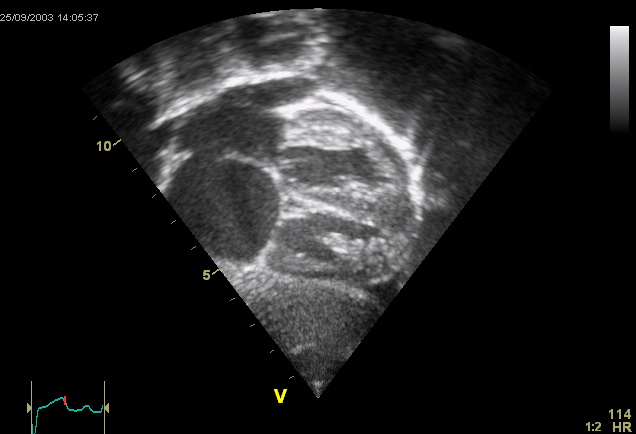
Echocardiogram showing an ostium secundum atrial septal defect (Wikimedia Commons, public domain)Widely fixed split S2, pulmonary flow murmur. Echo with agitated saline (bubble study) confirms shunt. TEE preferred for procedural planning.

Closure Indications

Class I: symptomatic with net L→R shunt (Qp:Qs >1.5) and no severe pulmonary vascular disease. RV enlargement even without symptoms is an indication.

PFO

25% of adults. Closure for cryptogenic stroke in select patients <60 with high-risk anatomy (atrial septal aneurysm, large shunt). RESPECT, CLOSE, REDUCE support closure over medical therapy.

Closure Criteria and Modalities

Secundum ASDs with favorable anatomy are typically closed percutaneously with a septal occluder device, while sinus venosus, primum, and coronary sinus defects require surgical repair. Closure is generally indicated for a hemodynamically significant shunt with evidence of right heart enlargement, even in an otherwise asymptomatic adult.
Clinical pearls
·
Sinus venosus ASDs are frequently missed on TTE — always image the SVC-RA junction on TEE if suspected.
·
Bubble study: agitated saline injected during Valsalva release — right-to-left shunt should appear within 3 cardiac cycles for intracardiac shunt.
·
A Qp:Qs ratio greater than 1.5:1, or evidence of RV volume overload on imaging, is the generally accepted threshold for hemodynamically significant shunting warranting closure.
·
Primum ASDs are part of the atrioventricular septal defect spectrum, are associated with a cleft mitral valve, and require surgical (not device) repair.
·
Even after technically successful closure, atrial arrhythmias remain common if closure is delayed past age 40, reflecting pre-existing atrial remodeling.
Related guidelines